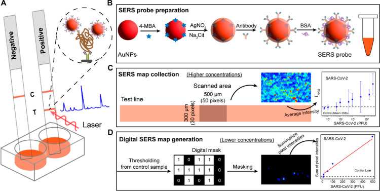
Figure 1.
Illustrative overview of the SERS–LFT dipstick for rapid SARS-CoV-2 detection. (A) SERS–LFT assay setup, involving sample mixing with an antibody-functionalized SERS probe and running buffer within a 96-well plate, followed by SERS spectra collection at the test line; (B) process of constructing the SERS probe, entailing the functionalization of the core–shell nanoparticle with Raman reporter and capture antibody; (C) 2D SERS mapping utilized for viral quantification at higher concentrations using average intensity measurements; and (D) 2D digital SERS mapping employed for quantifying lower viral concentrations based on the summation of pixel intensities.

