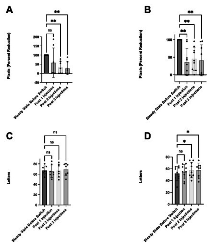
Figure 3. Comparing anatomy and function between fluid subtypes. The data has been represented in mean percent reduction +/- SD in TFA at one, two, and three injections relative to steady state in (A) eyes with SRF (n=7) and (B) eyes with IRF (n=9). The data has been represented in mean change in letters +/- SD at one, two, and three injections relative to baseline in (C) eyes with SRF (n=7) and (D) eyes with IRF (n=9).
*p<0.05, **p<0.01
IRF: intraretinal fluid, PED: pigment epithelial detachment, SRF: subretinal fluid, TFA: total fluid area reduction

