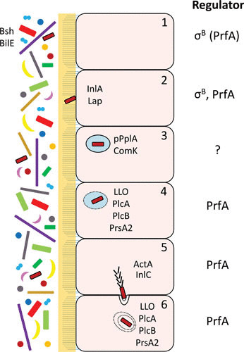
FIGURE 1.

Schematic overview of L. monocytogenes colonization of the intestine by pathways controlled by σB and/or PrfA. The human microbiota is depicted to the left, lying beside the microvilli-containing epithelial cells (1). Survival of L. monocytogenes in the gut is mediated by different factors, among them Bsh and BilE (2). The bacterium adheres to cells using different adhesins (e.g., InlA and Lap) (3). Once inside the phagosome, the pPplA and ComK systems become activated, facilitating phagosomal escape (4). Different PrfA-regulated factors mediate lysis of the phagosome (5). Through recruitment of the Arp2/3 complex, ActA allows polymerization of actin at the pole of the bacterium, driving it through the cytoplasm. The entry of L. monocytogenes to adjacent cells requires InlC, which relieves cortical tension (6). The escape from the double membrane vacuole requires the action of LLO, PlcA, and PlcB. Regulators (σB or PrfA) involved in the different regulatory steps during intestinal passage of L. monocytogenes are shown at the right.
