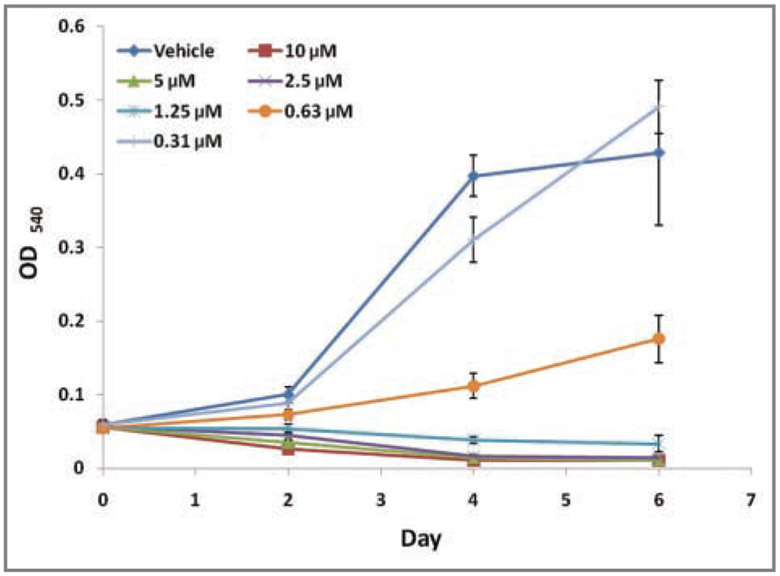
Figure 3—
Growth curves for SCCF1 cells in the presence of various concentrations of tepoxalin or a vehicle control (DMSO) by use of MTT assays. Optical density readings at a wavelength of 540 nm were obtained via spectophotometry at day 0 (prior to exposure to tepoxalin) and every other day thereafter during serial dilution treatment with tepoxalin or vehicle control. At day 6, all concentrations of tepoxalin (0.625 to 10μM) resulted in significant (P < 0.05) decreases in cell proliferation, compared with findings for vehicle control–treated cells.

