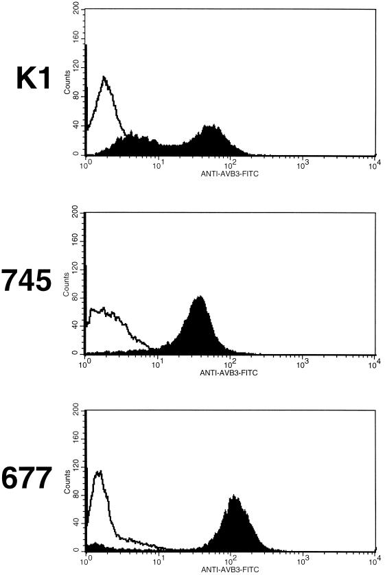
FIG. 2.
Analysis of CHO cells transfected with αvβ3 cDNAs. CHO-K1 cells and the two GAG-deficient mutants pgsA-745 and pgsD-677 were transfected with αvβ3 cDNAs and selected as described in Materials and Methods. After two to three passages, transfected and nontransfected cells were incubated with MAb LM609 for 30 min at 4°C in phosphate-buffered saline, washed, and incubated with fluorescein isothiocyanate (FITC)-labeled goat anti-mouse IgG for an additional 30 min at 4°C. After being washed with phosphate-buffered saline, cells were analyzed on a Becton Dickson FACSCaliber analyzer. Nontransfected cells are represented by open curves, and transfected cells are represented by closed curves.

