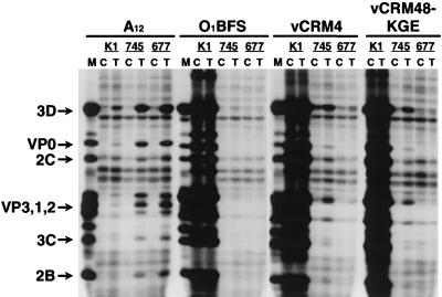
FIG. 3.
Analysis of viral proteins synthesized in FMDV-infected CHO cells transfected with αvβ3 cDNAs. Transfected (T) or nontransfected (C) CHO-K1, pgsA-745, or pgsD-677 cells were infected with FMDV type A12, O1BFS, vCRM4, or vCRM48-KGE at an MOI of 10 PFU/cell. Cells were labeled between 3 and 24 h after infection with [35S]methionine, and extracts were analyzed by RIP and SDS-PAGE as described in Materials and Methods. Viral proteins synthesized in infected and labeled BHK-21 cells are included as markers (M), and the positions of major viral proteins are indicated on the left.

