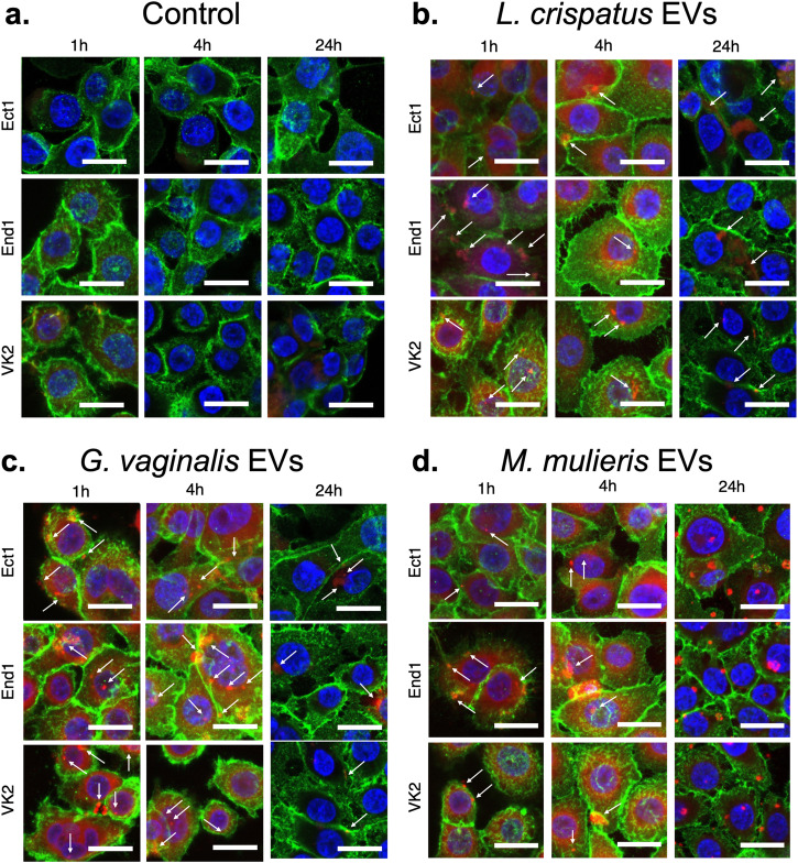
Fig. 3. Uptake of L. crispatus, G. vaginalis, and M. mulieris bEVs by cervical and vaginal epithelial cells over 24 h.
bEV preparations from (a) NYC culture medium (control), (b) L. crispatus, (c) G. vaginalis, and (d) M. mulieris were labeled with rhodamine B isothiocyanate and observed in the cytoplasm of ectocervical (Ect1), endocervical (End1), and vaginal epithelial (VK2) cells after 1, 4, and 24 h of incubation. All scale bars are 20 μm.

