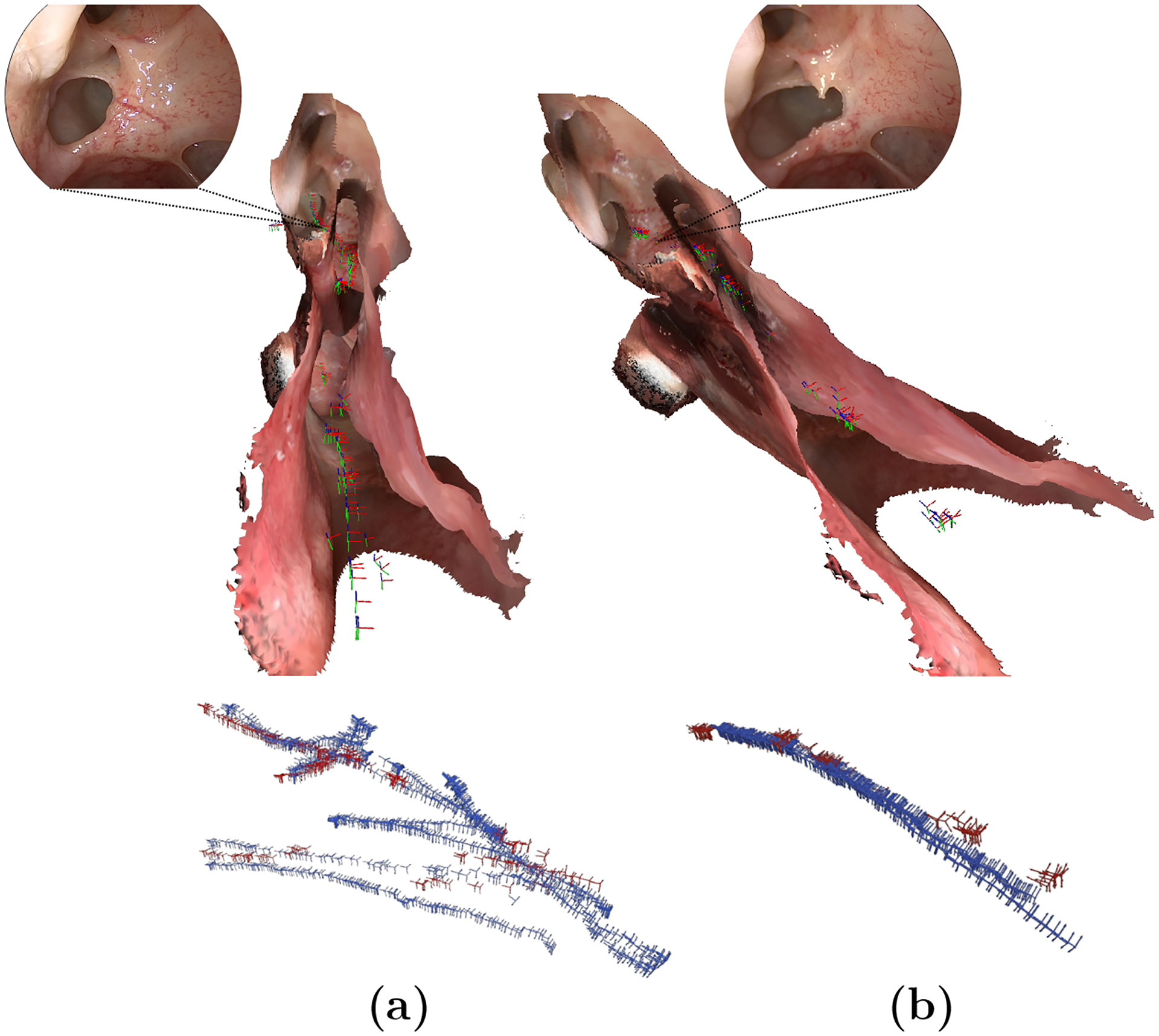
Fig. 2.

Spatial distribution of valid cameras (red) w.r.t its ground-truth trajectory (blue) and 3D anatomical model. (a) and (b) correspond to sequences of Undisturbed Anatomy and Progression Step # 1 of Subject # 1, respectively. A comparative frame of the operated region is also presented. Best viewed in color.
