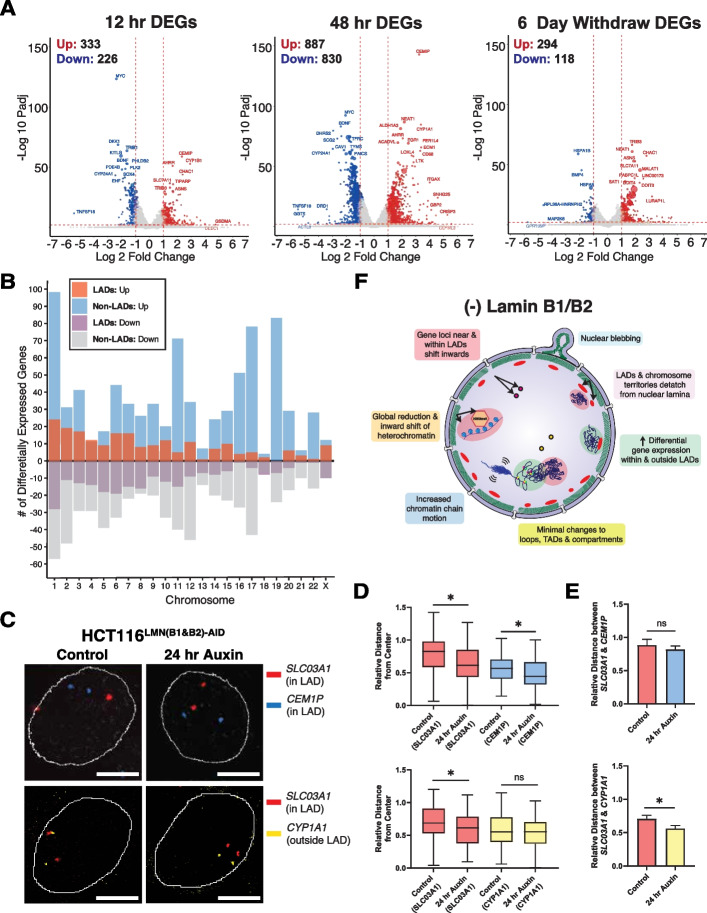
Fig. 8.
B-type lamin loss induces differential gene expression within and outside of LADs. A Volcano plots of DEGs after 12 h of auxin treatment, 48 h of auxin treatment, and 6 days of auxin withdrawal in HCT116LMN(B1&B2)−AID cells (adjusted P value < 0.01 and absolute log fold change > 1). B Bar plot of DEGs within or outside of LADs for each chromosome across all treatment time points. C Representative FISH images of gene loci within or outside of LADs in HCT116LMN(B1&B2)−AID cells. Scale bar = 5 μm. Data are representative of two independent biological replicates (N = 2). D Box plots of relative distances of SLC03A1, CEM1P, and CYP1A foci to the nuclear center (Control SLC03A1 (top) n = 50; Auxin SLC03A1 (top); n = 70; Control CEM1P n = 66; Auxin CEM1P n = 70; Control SLC03A1 (bottom) n = 116; Auxin SLC03A1 (bottom) n = 110; Control CYP1A1 n = 115; Auxin CYP1A1 n = 111)). The line within each box represents the mean; the outer edges of the box are the 25th and 75th percentiles and the whiskers extend to the minimum and maximum values. E Bar plots showing the relative distances between SLC03A1 and CEM1P foci and SLC03A1 and CYP1A1 foci (Mean + SEM). (SLC03A1 & CEM1P (Control n = 39; Auxin n = 57), SLC03A1 & CYP1A1 (Control n = 78; Auxin n = 57)). For D, E, data was compiled from two independent biological replicates (N = 2). Significance was calculated by Mann–Whitney test (* < 0.05). F Proposed model of the effect of Lamin B1 and B2 degradation on chromatin organization. Loss of B-type lamins induces nuclear blebbing and stalled cell cycle progression, indicating their structural and functional importance as components of the nuclear periphery. Although mid-range chromatin folding (e.g., TAD # and size, A/B compartmentalization, and contact probabilities in and outside LADs) is preserved, loss of these proteins promotes increased chromatin mobility along with an inward shift of chromosome territories (e.g., Chr. 1 and 2), heterochromatin-associated domains (e.g., LADs), and gene loci (red and yellow circles), especially at the nuclear periphery. The resulting genome-wide transcriptional changes both within and outside of LADs may be a direct consequence of heterochromatin redistribution and/or gene repositioning in relation to LADs upon the loss of structural constraints at the nuclear periphery

