Figure 2.

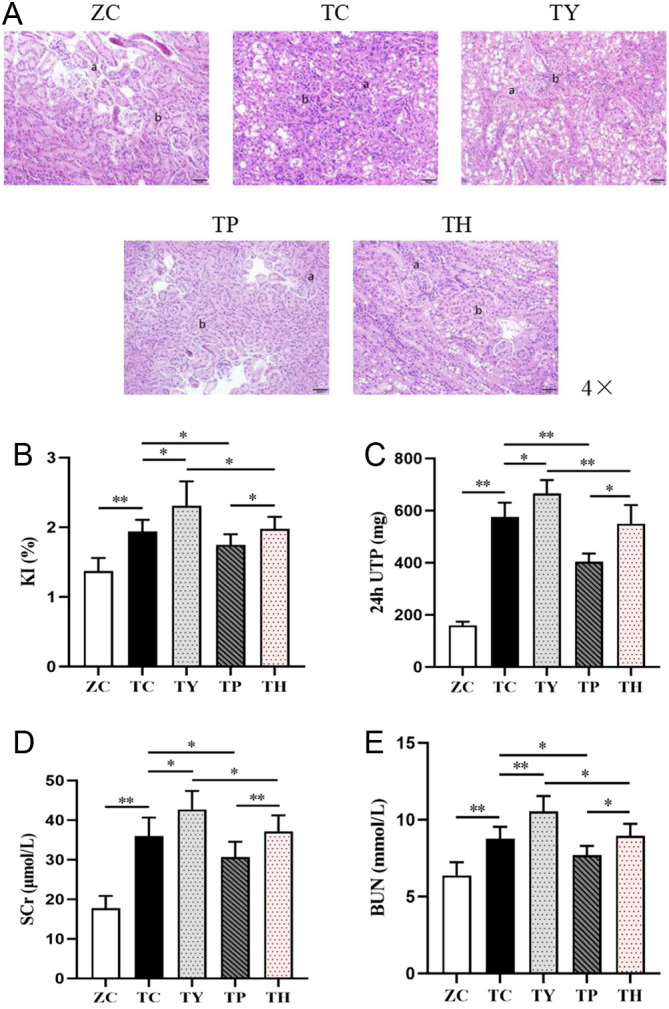
Renal injury in each group. (A) Representative images of H&E staining. Bar: 50 μm. (B, C, D, E) Expression levels of KI, 24UTP, SCr and BUN. (a) Glomerulus. (b) Renal interstitium. *P < 0.05, **P < 0.01.

Renal injury in each group. (A) Representative images of H&E staining. Bar: 50 μm. (B, C, D, E) Expression levels of KI, 24UTP, SCr and BUN. (a) Glomerulus. (b) Renal interstitium. *P < 0.05, **P < 0.01.