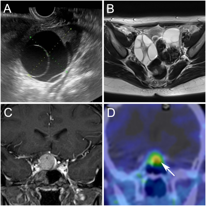
Figure 1.

Preoperative imaging findings of ovary and pituitary lesions. (A) Transvaginal ultrasound showed a large septated cystic ovary. (B) Pelvic MRI (axial T2-weighted image) showed bilateral multi-cystic ovaries. (C) preoperative pituitary MRI (coronal postcontrast T1-weighted image) revealed a mass lesion with suprasellar extension. (D) Preoperative 111In-pentetreotide scintigraphy showed increased tracer uptake in the pituitary lesion (arrow).

 This work is licensed under a