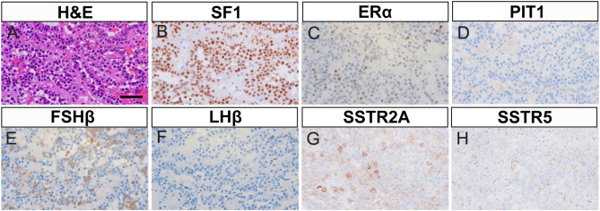
Figure 2.
Histopathology of resected pituitary tumor. High-magnification images of the resected pituitary tumor are shown (40× objective lens). (A) Hematoxylin and eosin staining (H&E), scale bar 50 µm. (B–D) Immunohistochemistry (IHC) for pituitary transcription factors. (B) Steroidogenic factor 1 (SF1). (C) Estrogen receptor alpha (ERα). (D) Pituitary-specific positive transcription factor 1 (PIT1). (E and F) IHC for gonadotropins. (E) Follicle-stimulating hormone subunit beta (FSHβ). (F) Luteinizing hormone subunit beta (LHβ). (G and H) IHC for somatostatin receptor (SSTR) subtypes. (G) SSTR2A. (H) SSTR5.

 This work is licensed under a