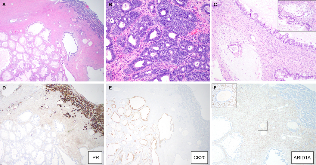
Figure 2:

Mixed gastro-intestinal type mucinous and endometrioid carcinoma (MO-1). (A) Low-magnification image showing spatial relationship of both components, with scattered benign cortical inclusion cysts; (B) endometrioid carcinoma component, (C) invasive glands floating in pools of mucin in a background of mucinous borderline tumor (inset: another invasive focus). Immunohistochemical stains for (D) PR, (E) CK20 and (F) ARID1A (inset: high magnification of boxed area highlights loss of ARID1A expression in the cortical inclusion cyst epithelium).
