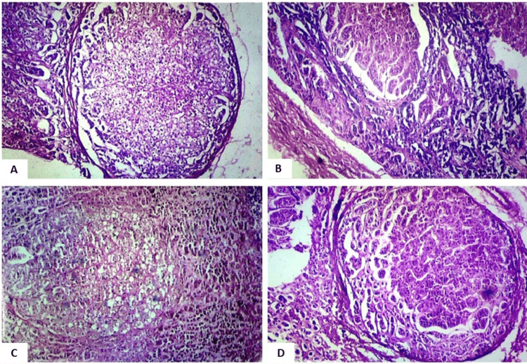
Figure 2. Photomicrograph showing nodule formation (regions of importance have been focused on in the field).
A. Nodule external to the capsule, magnified 40x (H&E stain).
B. Nodular formation in the zona glomerulosa, magnified 40x (H&E stain).
C. Nodular formation in the zona fasciculata, magnified 40x (H&E stain).
D. Nodular formation in the zona reticularis with brown lipofuscin pigments in cells, magnified 40x (H&E stain).

