
Figure 3.
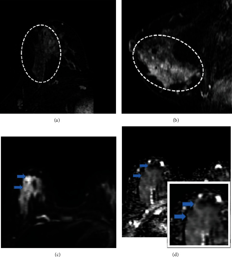
(a, b) Axial and sagittal contrast-enhanced breast MRI images in a 47-year-old female with pathology-proven invasive ductal carcinoma and ductal carcinoma in situ, depicting segmentally distributed clumped nonmass enhancement (dashed circle) in the right breast. (c, d) DWI and ADC images show heterogenous restriction which is better demonstrated in the inset image.