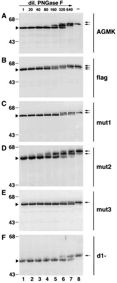
FIG. 4.
Partial PNGase F digestion of havcr-1 mutants. Cytoplasmic extracts of AGMK (A), flag (B), mut1 (C), mut2 (D), mut3 (E), and d1− (F) cells were prepared in RSB–1% NP-40. Aliquots containing 20 to 25 μg of total protein of each cytoplasmic extract were treated with undiluted PNGase F (500 U) (lanes 1), treated with twofold dilutions of PNGase F from 1:20 to 1:640 (lanes 2 to 7), or mock treated (lanes 8). After treatment, proteins were separated by SDS–12.5% polyacrylamide gel electrophoresis, transferred to PVDF membranes, and probed with rabbit anti-GST2 Ab directed against the TSP-rich region of havcr-1. Arrowheads point to completely N-deglycosylated forms of the receptors. Arrows point to N-glycosylated forms of the receptors expressed in the different cell lines. The positions of prestained molecular mass markers and their sizes in kilodaltons are shown on the left. dil., dilution.

