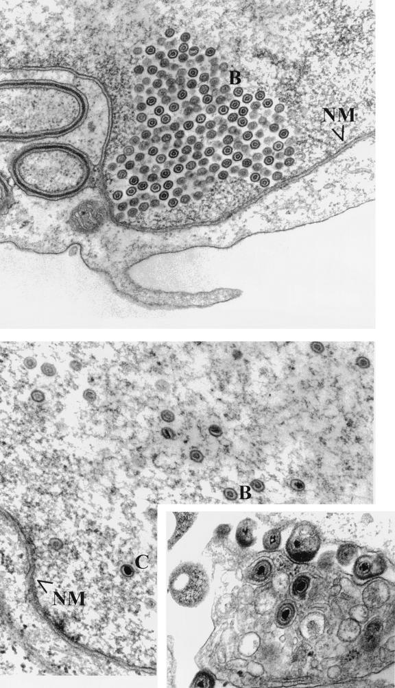
FIG. 5.
Scanned digital images of electron micrographs. Vero (top) and G5 (bottom) cells were fixed 14 h after infection with HSV-1(UL17-stop). The inset shows an image of the cytoplasm and extracellular space of G5 cells infected with HSV-1(UL17-stop). NM delineates the nuclear membrane; B and C indicate B and C capsids, respectively.

