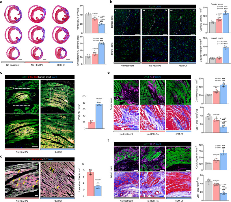
Fig. 8. Enhanced cardiac regeneration by human cardiac tissue transplantation in a rat model of myocardial infarction.
a Representative images of Masson’s trichrome (MT) staining of ischemic hearts harvested 4 weeks after transplantation and quantification of the percentage ratio of fibrotic and viable myocardium (scale bars = 2 mm, N = 5, biological replicates, *p < 0.05 and ***p < 0.001 versus No treatment group, ###p < 0.001 versus No HEM-Ps group). b Representative images of capillary staining for CD31 (green) (scale bars = 200 μm) and quantification of capillary density in the infarct zone (IZ) and the border zone (BZ) 4 weeks post-transplantation (N = 5, biological replicates, *p < 0.05 and ***p < 0.001 versus No treatment group, ###p < 0.001 versus No HEM-Ps group). c Representative immunofluorescent images comparing retention rates of transplanted RFP+ cardiomyocytes (CMs) in each group (total CMs stained with cTnT [green] and human CMs stained with human cTnT [white] after 4 weeks; scale bars = 100 μm), and quantification of engrafted human induced pluripotent stem cell (hiPSC)-derived CMs in ischemic hearts (N = 5, biological replicates, ***p < 0.001). d Representative immunofluorescent images of gap junctions expressed in transplanted RFP+ CMs (gap junctions stained with CX43 [green] and total CMs stained with cTnT [white]) 4 weeks post-transplantation. Yellow arrows indicate lateralized gap junctions (scale bars = 100 μm). The quantification graph shows the ratio of lateralized gap junctions to localized CX43 areas (N = 5, biological replicates, ***p < 0.001). Representative images of total CMs stained with cTnT (green) and denatured collagen stained with collagen hybridizing peptide (CHP, violet) on e the border zone and f the infarct zone after 4 weeks and their quantification data (scale bars = 100 μm, N = 5, biological replicates, *p < 0.05, **p < 0.01, and ***p < 0.001 versus No treatment group, ###p < 0.001 versus No HEM-Ps group). Cardiac tissues prepared with hiPSC-derived RFP+ CMs, cardiac fibroblasts (CFs), and human umbilical vein endothelial cells (HUVECs) were cultured under each condition for 9 days and used for transplantation. Data are presented as means ± S.D. Statistical significance was determined using unpaired two-sided Student’s t-tests in (c and d) and one-way ANOVA with Tukey’s multiple comparisons tests in (a, b, e, and f).

