Fig. 1.
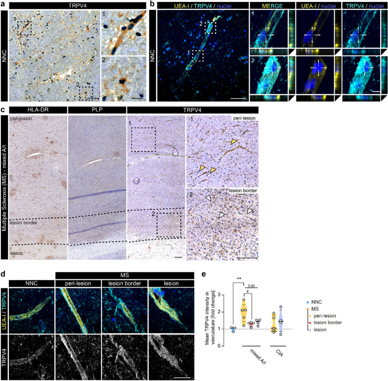
Vascular TRPV4 expression is increased in areas around inflammatory MS lesions. a Representative image of TRPV4 immunoreactivity in NNC WM tissue; scale bar: 100 µm. Panels highlight morphologically different types of TRPV4+ cells including glial and vascular cells; scale bar: 25 µm. b Representative confocal image of UEA-I (endothelial marker) and TRPV4 immunoreactivity in NNC; scale bar: 50 µm. Panels demonstrate TRPV4-UEA-I co-localization; scale bar: 5 µm. c Representative images of HLA-DR, PLP, and TRPV4 immunoreactivity in mixed active/inactive (A/I) WM lesion tissue. Overview TRPV4 image shows the staining pattern at low magnification of peri-lesional, lesion border, and lesion tissue respectively (dotted line indicates areas, squares indicate location of panels); scale bar: 200 µm. Panels demonstrate TRPV4 staining in peri-lesional (orange arrowheads) and lesion border tissue (white arrowheads). d Representative confocal images of TRPV4-UEA-I immunoreactivity in NNC and MS cases; scale bar: 50 µm. e Quantification of TRPV4 mean fluorescent intensity measured within the UEA-I signal (endothelium) in NNC (N cases = 3) and MS (mixed A/I): N cases = 3, N lesions = 5; chronic inactive (CIA): N cases = 4, N lesions = 5). Statistical analysis was performed using one-way ANOVA with Dunnett’s multiple comparisons test, followed by paired one-way ANOVA analysis within the MS cases (#). Violin plots show median ± quartiles (*p < 0.05, **p < 0.01; #p < 0.05)

