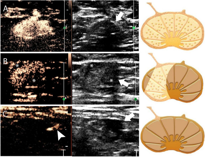
Fig. 1.
Enhancement patterns of the SLNs on contrast-enhanced lymphatic US. From left to right: the enhanced US image; the grayscale US image at the dual display; and the schematic illustration of enhanced and grayscale US images. A Pattern I, homogeneous enhancement. The echogenic contrast agent is evenly distributed within the SLN with a thin hypoechoic cortex (arrows) on the grayscale US. B Pattern II, inhomogeneous enhancement. The uneven distribution of the contrast agent with two filling defect areas (*) of the SLN is seen correlating to the focal eccentric cortical thickening (arrows) on grayscale US. C Pattern III, no enhancement. There is an enhanced lymphatic vessel (arrowhead), but no contrast agent accumulation is visible inside the SLN. The SLN shows focal eccentric cortical thickening on grayscale US (arrows). SLN sentinel lymph node

