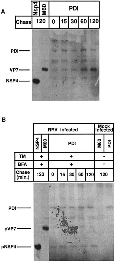
FIG. 5.
Cells infected with RRV (MOI of 10) were mock (A) or BFA (2 μg/ml) treated (B) at 1 hpi. At 5 hpi, TM (2 μg/ml) was added to the medium of the monolayers (as indicated) and was maintained through the experiment. At 7 hpi, cells were starved for 1 h in methionine-cysteine-free media and then metabolically labeled (200 μCi) for 15 min. Labeled proteins were chased with Eagle’s MEM supplemented with 1 mM cycloheximide and 10 mM methionine for the lengths of time indicated over the lanes. At the end of the pulse or chase, monolayers were incubated with ice-cold PBS with 40 mM NEM for 2 min, and cells were harvested in gentle lysis buffer. The cell lysates were immunoprecipitated with MAbs to VP7 and NSP4 and rabbit polyclonal antibodies to PDI.

