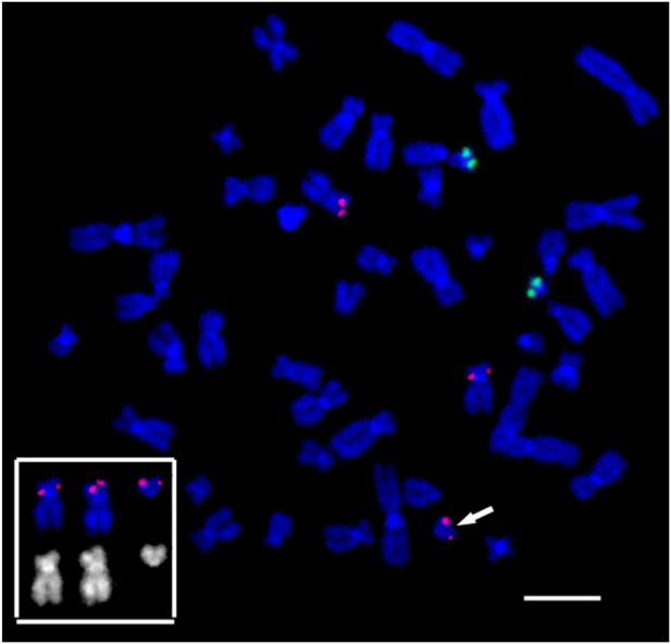
FIGURE 10.

Suppression fluorescence in situ hybridization of locus-specific DNA probes on the patient’s metaphase chromosomes. FISH TEL/AML1 Translation, Dual Fusion Probe (Cytocell, Cambridge, UK). TEL (12p13.2)–red, AML1 (21q22.12)–green. Chromosome 12 and marker 12, bearing a red signal in the 12p13.2 locus, and their inverted DAPI banding are shown at the bottom left. The arrow points to the small supernumerary marker chromosome 12 found in the patient’s karyotype. General chromosome staining with DAPI (blue signal). Scale bar 50 µm.
