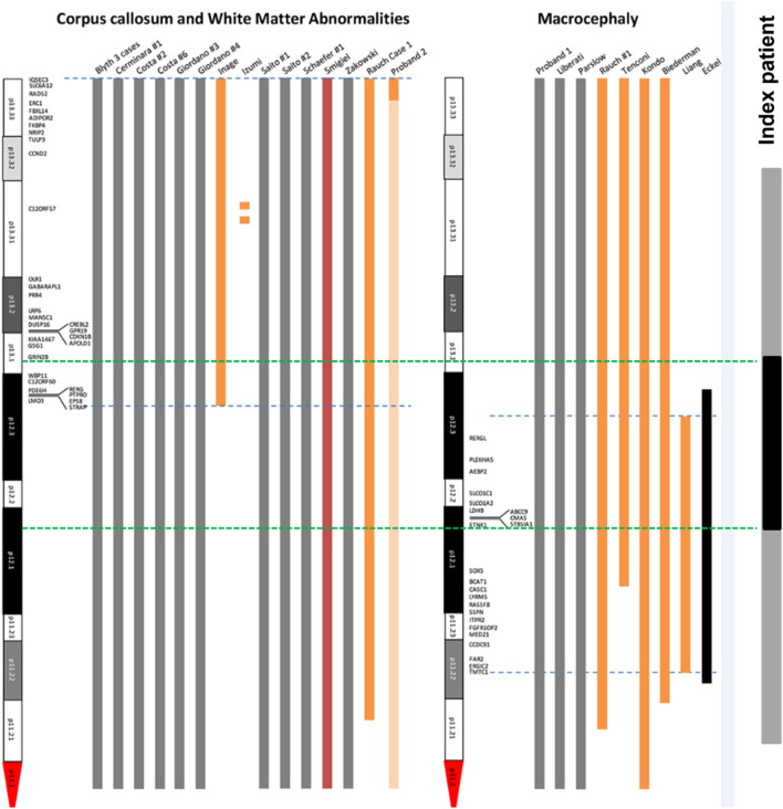
FIGURE 11.
Mapping of candidate genes of structural brain abnormalities in patients with PKS. The figure is based and modified from the original map by Poulton et al. (2018) (Poulton et al., 2018). Blue dotted lines are the boundaries of localization of genes associated with abnormalities of corpus collosum and white matter (left) and macrocephaly (right). Green dotted lines are the boundaries of duplicated 12p13.1-p12.1 region (shown in black) within the sSMC(12) (shown in grey) in our patient.

