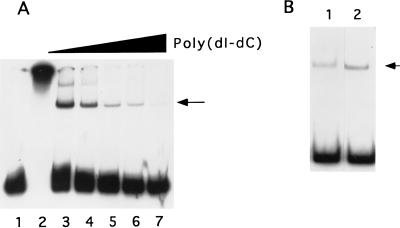
FIG. 1.
(A) Detection of late promoter binding activity in Ni-agarose-purified protein. Extracts from vaccinia virus-infected cells were chromatographed on Ni-agarose and assayed for binding to the 32P-labeled 120-nucleotide 11-kDa promoter probe. Lane 1 contains probe alone; lanes 2 to 7 contain probe plus 200 ng of protein. Poly(dI-dC) was included in binding reactions at 0 (lane 2), 100 (lane 3), 200 (lane 4), 500 (lane 5), 1,000 (lane 6), and 2,000 ng (lane 7). Protein-DNA complexes were resolved by electrophoresis in a native gel and visualized by autoradiography. The location of the major protein-probe complex is indicated with an arrow. (B) Ni-agarose-purified protein from uninfected HeLa cells (lane 1) or vaccinia virus-infected cells (lane 2) was tested for binding to the 11-kDa promoter probe. The major protein-probe complex is indicated with an arrow.

