
Figure 1.
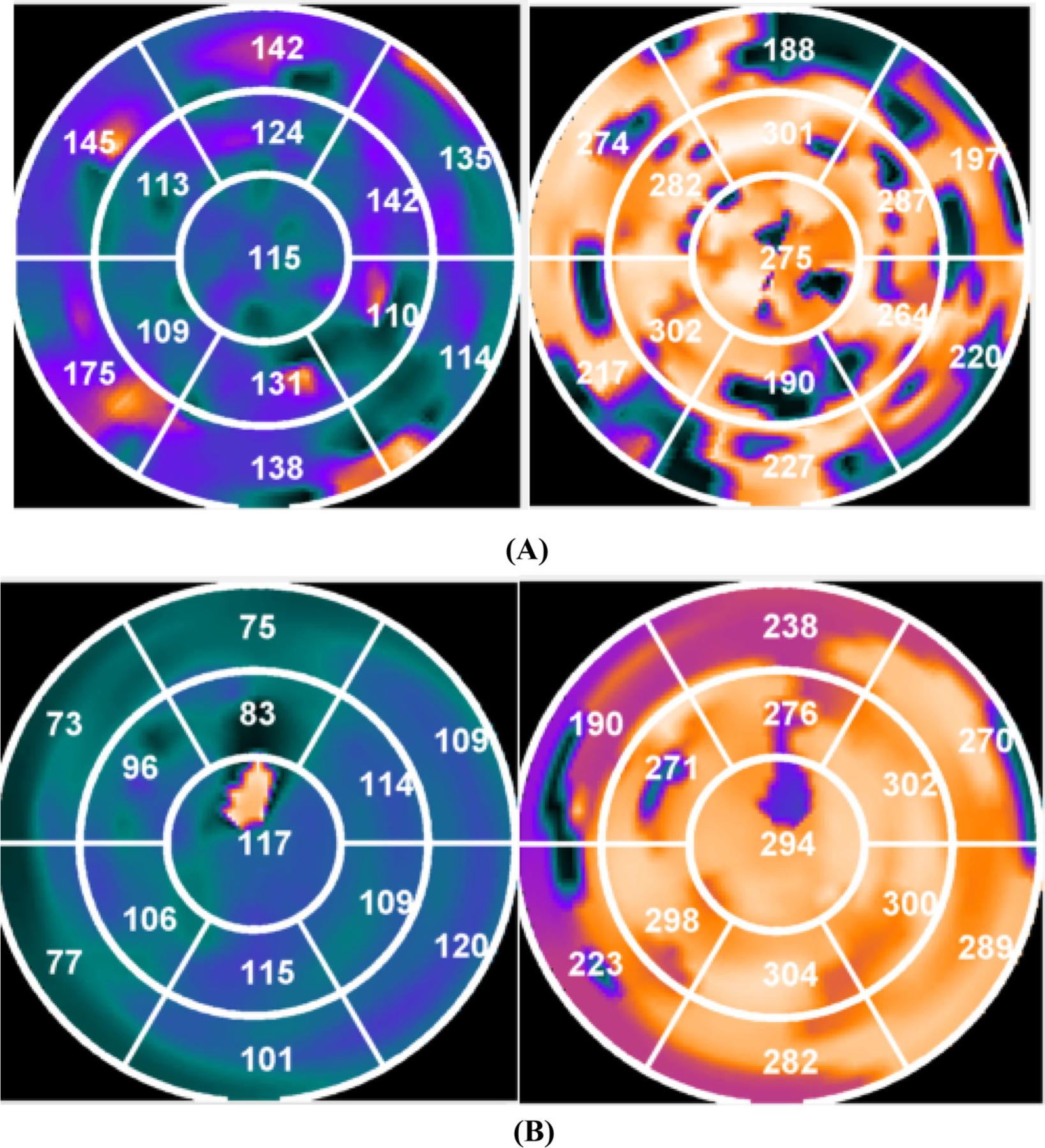
Polar maps showing systolic and diastolic dyssynchrony. (A) shows a patient who didn’t respond to CRT. The latest three contracting segments were in the septal and anterior wall and the latest three relaxing segments were in the septal, anterior, and anterolateral wall. The left ventricular lead was placed in the posterolateral wall. (B) shows a patient who responded to CRT. Both the latest three contracting and latest three relaxing segments were all in the posterolateral wall. The left ventricular lead was placed in the posterolateral wall.