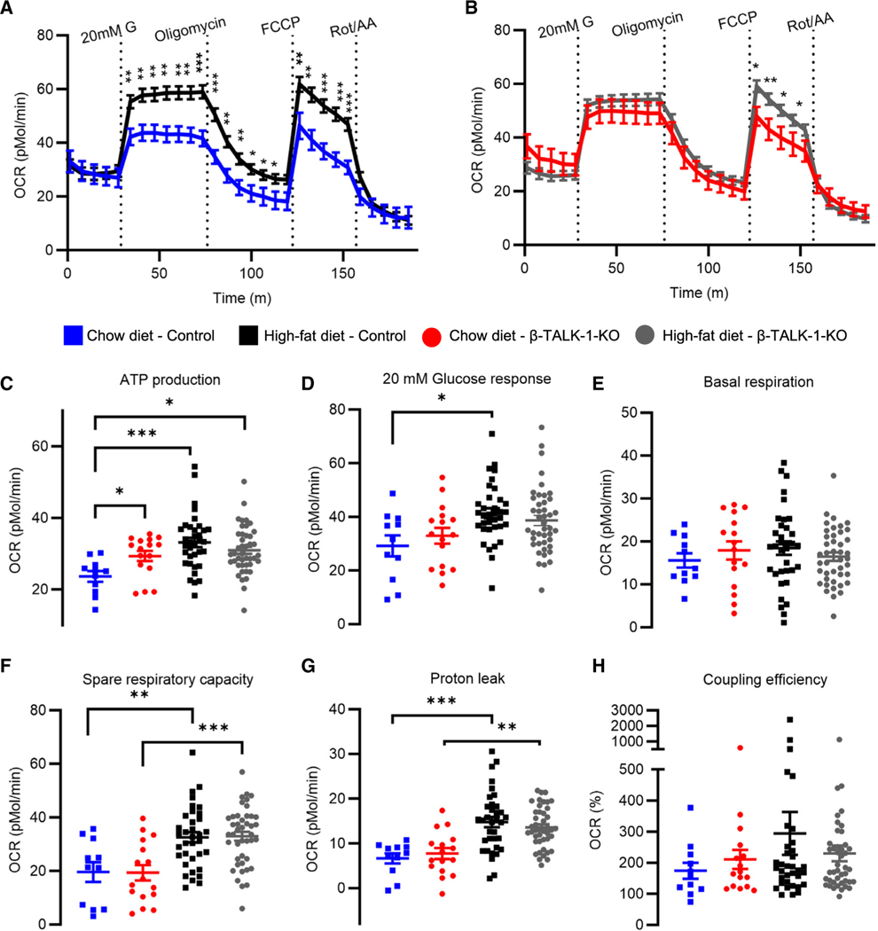
Figure 3. β-cell TALK-1 deficiency increases mitochondrial ATP production.
(A and B) Average OCR profiles measured in control islets (A) and β-TALK-1-KO islets (B) from mice fed either a chow diet or an HFD using the Agilent Seahorse XF Cell Mito Stress Test Kit. Islets were consecutively treated with 20 mM G, 4.5 μM oligomycin, 1 μM FCCP, and 2.5 μM rotenone/antimycin A (Rot/AA).
(C) ATP production rate was calculated by subtracting the minimal rate after oligomycin injection from the last rate measurement before oligomycin injection.
(D) Glucose-stimulated OCR response was calculated by subtracting basal respiration rate from maximal OCR in response to 20 mM G.
(E) Basal respiration rate was calculated by subtracting non-mitochondrial oxygen consumption from last rate measurement before first injection.
(F) Spare respiratory capacity is measured as the ratio of basal respiration to maximal respiration after FCCP injection (*100).
(G) Proton leak was calculated by subtracting non-mitochondrial oxygen consumption from minimum OCR after oligomycin addition.
(H) Coupling efficiency is the ratio of ATP-production coupled respiration to basal respiration (*100).
Data are represented as mean ± SEM. n = 11 wells from 3 control mice (chow diet), n = 16 wells from 3 β-TALK1-KO mice (chow diet), n = 35 wells from 4 control mice (HFD), and n = 43 wells from 7 β-TALK1-KO mice (HFD). Statistical significance was determined by one-way ANOVA, *p < 0.05 and **p < 0.01.

