
FIGURE 1.
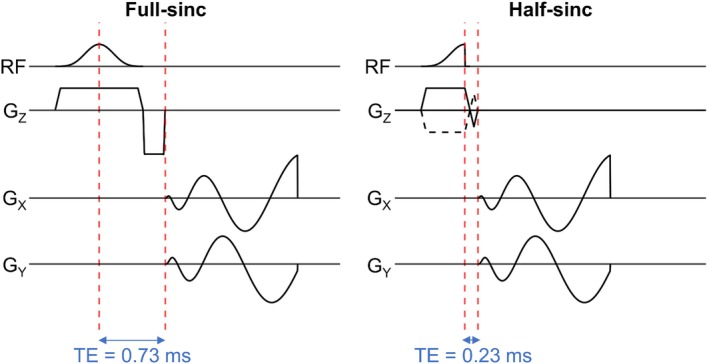
Pulse sequence diagrams for 2D sodium imaging with a center‐out spiral k‐space trajectory and a single‐lobe full‐sinc excitation pulse (left) or a half‐sinc excitation pulse (right). The TE is reduced from 0.73 ms with the full‐sinc pulse, and to 0.23 ms with the half‐sinc pulse; however, with the half‐sinc pulse it is necessary to acquire data across two acquisitions, with slice‐select gradients of opposite polarity. These two acquisitions are summed to give the same slice profile as the full‐sinc. GZ/GX/GY, magnetic field gradients applied along the Z, X, and Y axes, respectively, where Z is the slice‐select axis and X and Y lie in the plane of the imaging slice; RF, radiofrequency excitation pulse.