
Figure 1.
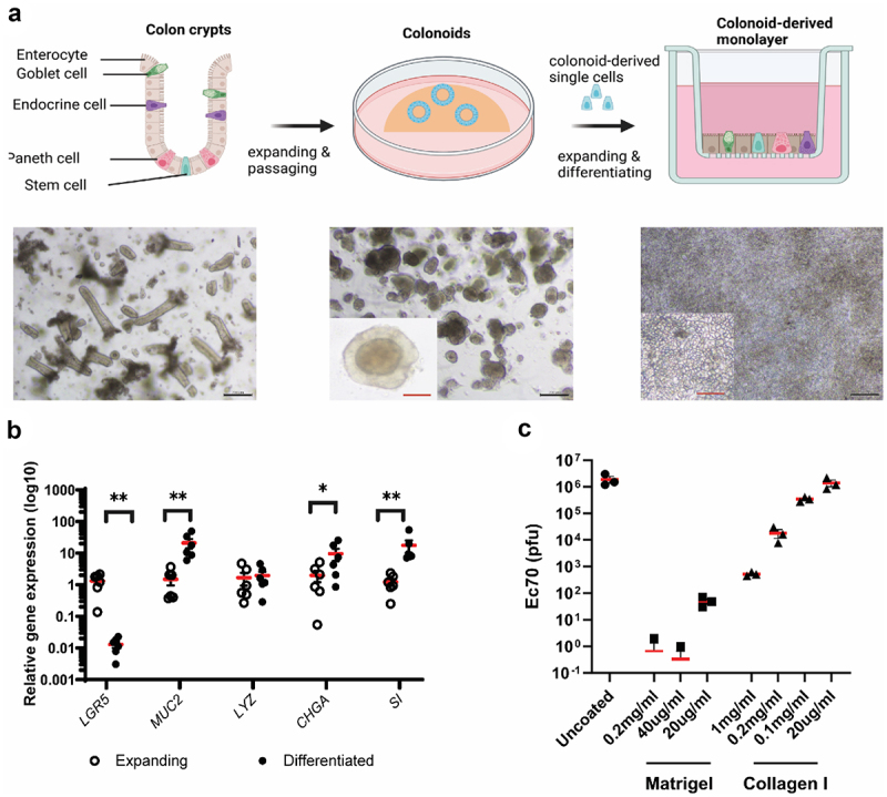
Development of colon-derived epithelial monolayer for phage translocation study.
a. Colon crypts (left) were isolated from colon biopsies and grown into 3D colonoids (centre) which were then dissociated into single cells and seeded on transwells to form a colon-derived epithelial monolayer (right). b. Expression of colon epithelial cell markers in expanding and differentiated layers. Markers: LRG5, stem cells; MUC2, Goblet cells; LYZ, Paneth cells; CHGA, endocrine cells; SI, enterocytes. Wilcoxon test, n=6, * p < .05, ** p < .01. c. Optimising transwell-coating substances to allow free diffusion of translocated phages. Black bar scale,200 nm; red bar scale, 50 nm.