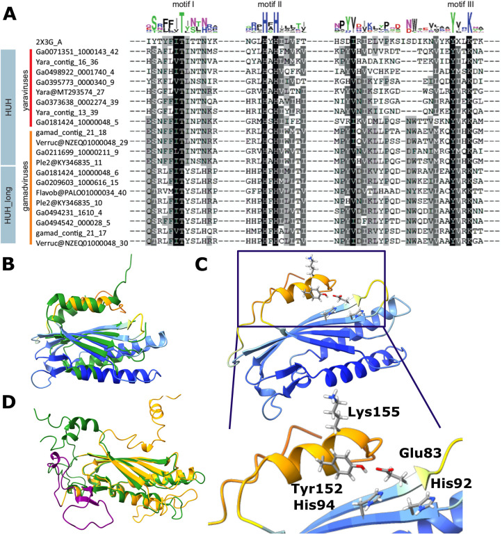
Figure 6. Sequence and structure conservation in the HUH endonucleases of mriyaviruses.
A, Alignment of the sequence segments of the HUH superfamily endonucleases containing the characteristic motifs I-III (N-terminal motif I consisting of hydrophobic residues, motif II with (HUH; H: Histidine, U: hydrophobic residue) and C-terminal motif III (Yx2-3K; Y: tyrosine, x: any residue, K: lysine, blue), where only the second tyrosine is present (compared to the full motif 3 YUxxYx2-3K, U: hydrophobic residue), are highlighted. B, A representative predicted structure of a mriyavirus HUH endonuclease superimposed with the crystal structure of protein ORF119 from Sulfolobus islandicus rod-shaped virus 1 (green, pdb 2X3G-A, z-score 7.7). Yaravirus HUH endonuclease (MT293574_27) colored by plddt score. C, Configuration of the catalytic amino acid residues of motif II and III in the predicted structure of the mriyavirus HUH endonuclease (Yaravirus MT293574_27, colored by plddt score). D, Superposition of the structural models of the two HUH endonuclease domains of gamadviruses (short, probably active: KY346835_11 (green, aa 31–224, aa1-30 unstructured, clipped off for representation), long: KY346835_10 (orange, aa 1353–1574 with additional inserted loop (purple) aa 104–1450).

