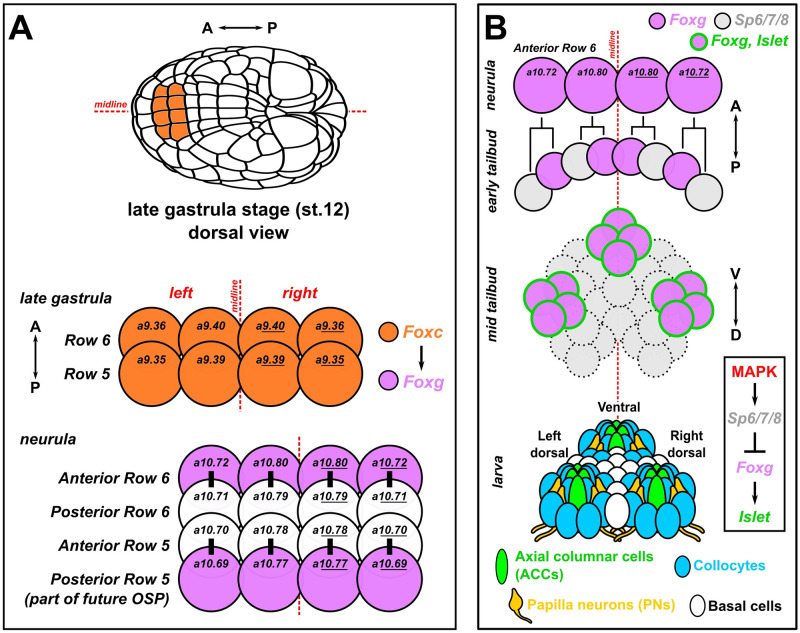
Fig 1. Development of the papillae of Ciona.
(A) Diagram showing the early cell lineages that give rise to the papillae. The papillae invariantly derive from Foxc+ cells in the anterior neural plate, more specifically the anterior daughter cells of “Row 6” of the neural plate, which activate Foxg downstream of Foxc. Foxg is also activated in the posterior daughter cells of “Row 5,” which go on to give rise to part of the OSP. Numbers in each cell indicate their invariant identity according to the Conklin cell lineage nomenclature. Black bars indicate sibling cells born from the same mother cell. (B) Diagram of what is currently known about the later lineage and fates of the Foxg+ “Anterior Row 6” cells shown in panel A. As the cells divide mediolaterally, some cells up-regulate Sp6/7/8 and down-regulate Foxg (gray cells). Those cells that maintain Foxg expression turn on Islet and coalesce as 3 clusters of cells (pink with green outline): 1 medial, more ventral cluster, and 2 left/right, more dorsal clusters. Later, these 3 clusters organize the territory into the 3 protruding papillae of the larva, which contains several cell types described in detail by TEM [9]. Dashed cell outlines indicate uncertain number/provenance of cells. A-P: anterior-posterior. D-V: dorsal-ventral. Lineages and gene networks are based mostly on: [21,22,86,87]. OSP, oral siphon primordium; TEM, transmission electron microscopy.

