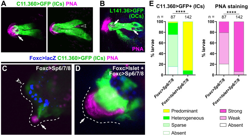
Fig 5. Both types of collocytes contribute to production of adhesive material.
(A) PNA-stained granules (pink) are seen in the hyaline cap and the apical tip of ICs (left panel, white arrow) in a C. intestinalis larva labeled by the C. intestinalis C11.360>Unc-76::GFP reporter (green). PNA-stained granules are also seen in cells not labeled by the IC reporter (right subpanel, hollow arrowhead), suggesting they are localized in a different cell type. Left and right subpanels are from different focal planes of the same papilla. (B) OCs labeled with C. robusta L141.36>Unc-76::GFP (green) in a C. robusta larva, with PNA-stained granules (pink) in both apical and basal positions within the cell (white arrows). DAPI in blue. (C) PNA staining (pink) in C. robusta upon overexpression of Sp6/7/8 alone, showing reduction of IC specification as assayed by C11.360>Unc-76::GFP expression (green). Weak PNA staining and GFP expression are still visible in some papillae (solid arrow), but not others (open arrowhead). (D) PNA staining (pink) and C11.360>Unc-76::GFP expression (green) in C. robusta upon overexpression of both Islet and Sp6/7/8, showing expansion of IC fate in a single large papilla (arrow). PNA staining is similarly expanded over the entire IC cluster, confirming that ICs produce the adhesive glue. Foxc>lacZ expression (β-galactosidase immunostaining) shown in blue in both C and D. (E) Scoring of larvae represented in panels C and D, averaged across duplicates. Weak PNA staining is observed upon partial suppression of IC fate, but strong PNA staining is seen upon expansion of supernumerary ICs, confirming that this cell type is one of the major contributors of PNA-positive adhesive glue. Total larvae (duplicate 1) or β-galactosidase+ larvae (duplicate 2) were scored. **** p < 0.0001 in both duplicates, as determined by chi-square test. See S4 Data for sample size, statistical test details, and for the data underlying the graphs. C. intestinalis raised to 20–22 hpf at 18 °C (~st. 28), C. robusta raised to 20 hpf at 20 °C (~st. 29). See Supplemental Movies for full confocal stacks and S6 Fig for single-channel images. IC, inner collocyte; OC, outer collocyte; PNA, peanut agglutinin.

