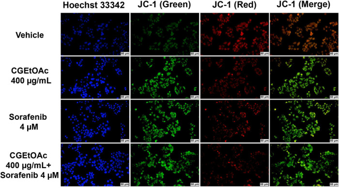
Fig 4. Determination of mitochondrial membrane potential (MMP) in HepG2 cells after exposure to CGEtOAc, sorafenib, and their combination for 24 h by labeling with 5,5,6,6′-tetrachloro-1,1′,3,3′ tetraethylbenzimidazoylcarbocyanine iodide (JC-1) and Hoechst 33342.
The images were detected using a fluorescent microscope with a magnification bar of 50 μM. Cells treated with 0.8% DMSO represented the vehicle control.

