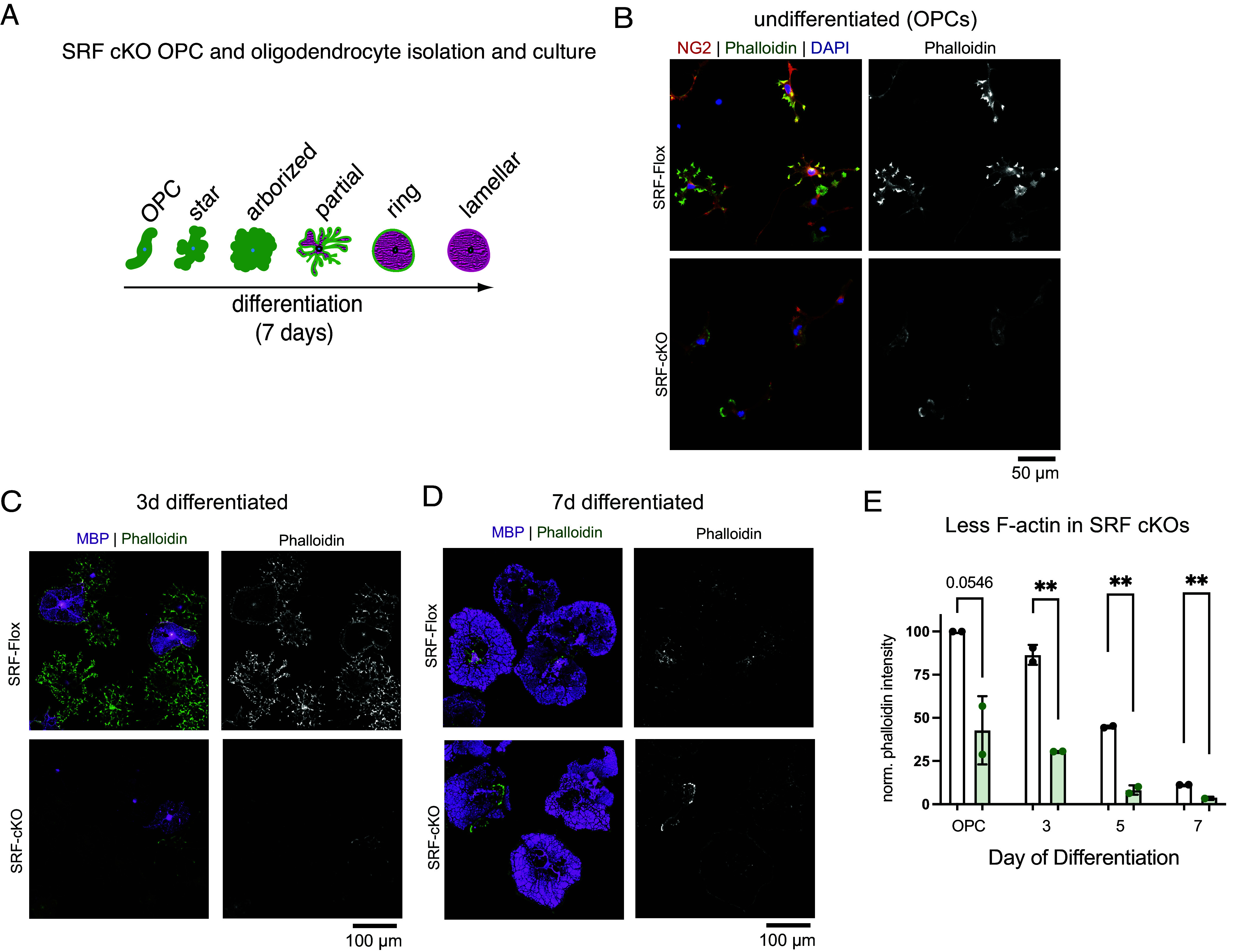
Fig. 5.

SRF regulates the oligodendrocyte cytoskeleton. (A) Diagram of OPC to oligodendrocyte morphology changes when differentiated in culture. (B) SRF-Flox and SRF-cKO OPCs stained for the OPC marker NG2 (red), phalloidin (green) and DAPI (blue). (Scale bar, 50 μm.) (C) SRF-Flox and SRF-cKO immature oligodendrocytes (3 d differentiated) stained for MBP (magenta), phalloidin (green), and DAPI (blue). (Scale bar, 100 μm.) (D) SRF-Flox and SRF-cKO mature oligodendrocytes (7 d differentiated) stained for MBP (magenta), phalloidin (green) and DAPI (blue). (Scale bar, 100 μm.) (E) Quantification of phalloidin intensity in SRF-Flox and SRF-cKO at different stages of differentiation. n = 2 preps. Unpaired t test; **P < 0.01.
