Fig. 5.
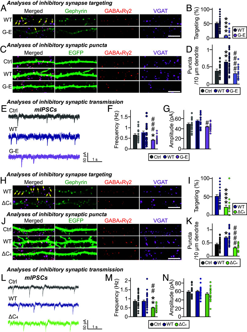
The importance of the C domain in inhibitory synapse formation. (A) Triple-immunofluorescence images of anti-GABAARγ2 (red), anti-VGAT (magenta) and anti-EGFP (green) antibodies in cultured hippocampal neurons transfected with EGFP-tagged gephyrin WT and G-E variant. Yellow arrows mark colocalization of gephyrin with both GABAARγ2 and VGAT puncta. (Scale bar, 10 μm.) (B) Quantification of inhibitory synaptic targeting of gephyrin variants in cultured hippocampal neurons (2–3 dendrites per transfected neuron were analyzed and group-averaged). (WT and G-E ****P < 0.0001). Error bars ± SEM. (C) Triple-immunofluorescence images of anti-GABAARγ2 (red), anti-VGAT (magenta) and anti-EGFP (green) antibodies in cultured hippocampal neurons transfected with EGFP alone or co-transfected with EGFP and EGFP-tagged gephyrin variants. (Scale bar, 10 μm.) (D) Effects of overexpression of gephyrin WT or the indicated variants on GABAARγ2+VGAT+ puncta density (2–3 dendrites per transfected neuron were analyzed and group averaged). (Ctrl and WT **P = 0.0017, WT and G-E ####P < 0.0001). Error bars ± SEM. (E–G) Representative traces and quantification of frequency and amplitude of mIPSCs recorded from hippocampal cultured neurons transfected with the indicated gephyrin variants. (F) Ctrl and G-E *P = 0.0258, WT and G-E ###P = 0.0004; (G) WT and G-E #P = 0.0351). Error bars ± SEM; Control, n = 19; WT, n = 21; G-E, n = 19. (H) Triple-immunofluorescence images of anti-GABAARγ2 (red), anti-VGAT (magenta) and anti-EGFP (green) antibodies in cultured hippocampal neurons transfected with EGFP-tagged gephyrin WT and ∆C4 variant. Yellow arrows mark colocalization of gephyrin with both GABAARγ2 and VGAT puncta. (Scale bar, 10 μm.) (I) Inhibitory synaptic targeting of gephyrin variants in cultured hippocampal neurons (2–3 dendrites per transfected neuron were analyzed and group-averaged). (WT and ∆C4 ****P < 0.0001). Error bars ± SEM. (J) Triple-immunofluorescence images of anti-GABAARγ2 (red), anti-VGAT (magenta) and anti-EGFP (green) antibodies in cultured hippocampal neurons transfected with EGFP alone or co-transfected with EGFP and EGFP-tagged gephyrin variants. (Scale bar, 10 μm.) (K) Effects of overexpression of gephyrin WT or the indicated variants on GABAARγ2+VGAT+ puncta density (2–3 dendrites per transfected neuron were analyzed and group averaged). (Ctrl and WT *P = 0.0365, WT and ΔC4 ####P < 0.0001). Error bars ± SEM. (L–N) Representative traces and quantification of frequency and amplitude of mIPSCs recorded from hippocampal cultured neurons transfected with the indicated gephyrin variants. [(M) Ctrl and ΔC4 **P = 0.0031, WT and ΔC4 ##P = 0.0015]. Error bars ± SEM; Control, n = 23; WT, n = 28; ΔC4, n = 22.

