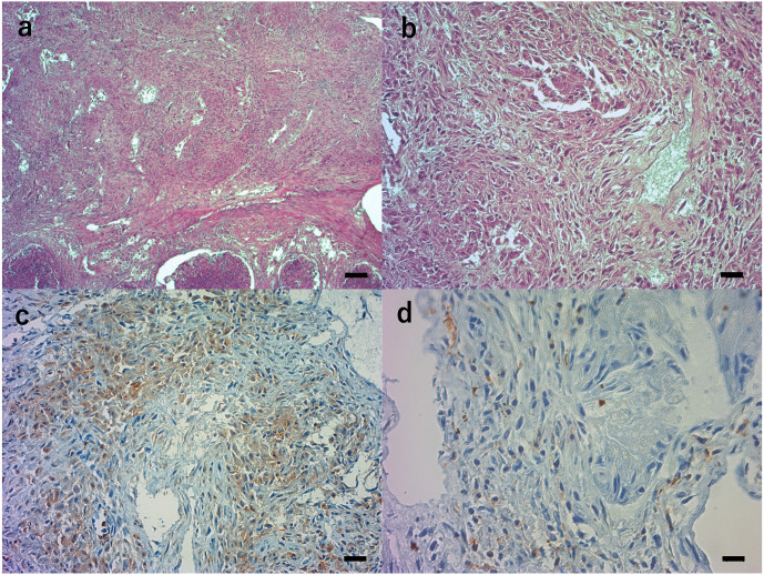
Fig. 2.
Histopathological examination of the left testis of the stranded rough-toothed dolphin. Tissue sections were stained with hematoxylin and eosin (a, b) or immunostained with primary antibodies against leukocytes (c, d). a and b. The parenchyma of the left testis was diffusely necrotic with granulomatous infections consisting of many macrophages, epitheloid cells, and some plasma cells, neutrophils and lymphocytes. Black bar: 100 μm (a), 20 μm (b). c. Many macrophages and epithelioid cells were positive with the lba1 polyclonal antibody. Black bar: 20 μm. d. Some lymphocytes were positive with the monoclonal antibody BLA36. Black bar: 10 μm.

