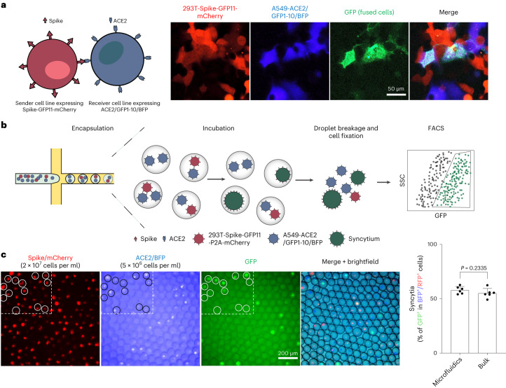
Fig. 1. A droplet microfluidics-based system for high-throughput screening of syncytium formation.
a, Co-culture of spike-expressing sender cells (mCherry+) and ACE2-expressing receiver cells (BFP+) resulted in syncytium formation (GFP+) due to cell–cell fusion. Scale bar, 50 μm. b, Workflow for cell encapsulation and incubation in droplets and the detection/collection of GFP+ syncytium (boxed). Side scatter (SSC) was used to identify the cell population. c, Representative images of sender cell–receiver cell fusion in droplets. A fusion rate of 57.9% ± 3.3% (data shown are mean ± s.d., n = 6 biological replicates, with P value from unpaired two-tailed Student’s t-test) was measured by FACS after 24 h of incubation in droplets, which is comparable to the bulk setting (55.2% ± 4.3%) in regular co-culture experiments without using droplet microfluidics. Red fluorescent protein (RFP) refers to the mCherry signal detected under FACS.

