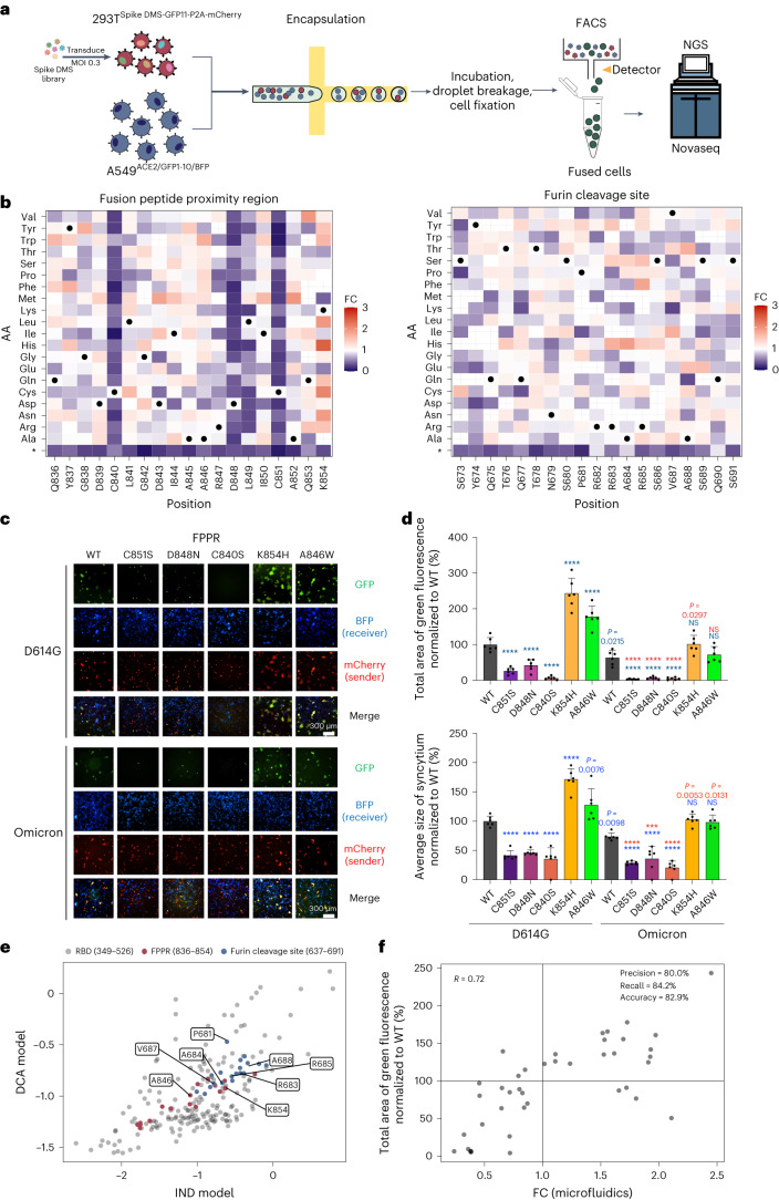
Fig. 2. Deep mutational scanning of SARS-CoV-2 spike’s syncytium-forming potential using the droplet microfluidics-based system.
a, Workflow for deep mutational scanning in sender cells using a droplet microfluidics-based system. GFP+ syncytia containing the fusion-competent spike variants are sorted out for NovaSeq-based sequencing. b, Heat maps depicting how all single mutations affect the syncytium-forming potential of spike’s FPPR and furin cleavage site regions. Squares are coloured by mutational effect (that is, FC) according to colour bars on the right, with red and purple indicating syncytium-enhancing and inhibiting substitutions, respectively. FC represents each variant’s relative abundance in the sorted GFP-positive cell pool vs the cell pool before mixing and is normalized to WT. Spike variants with increased syncytium-formation potential were enriched (with fold change >1), while those with decreased syncytium-formation potential were depleted (with fold change <1). Grey cross, mutations with no measurement; black dot, SARS-CoV-2 WT amino acid; *, stop codon. c, Validation of syncytium-enhancing and inhibiting mutations at the FPPR in WT D614G and Omicron spike. GFP-split complementation system assay was applied and representative images are shown. d, Quantification of the syncytium area (top) and average size of syncytium (bottom) for spike variants in c. Data shown are mean ± s.d.; n = 6 biological replicates. P values coloured in blue indicate statistical comparison between individual spike D614G and Omicron mutants to the D614G WT. P values coloured in red indicate statistical comparison between individual Omicron mutants to the omicron WT. Statistical significance was determined using one-way ANOVA. ***P < 0.001, ****P < 0.0001. e, Mutability score for individual position of spike protein studied in b and RBD. The number indicates the position of the amino acid on the spike protein. The letter indicates the wild-type amino acid on the position. f, Mutational effects on syncytium formation revealed by the pooled screen using the droplet microfluidics-based system and individual validation assay for the spike variants. Black dots, validated spike FPPR variants.

