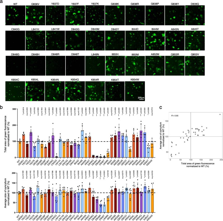
Extended Data Fig. 3. Individual validation of FPPR variants of Spike.
a) GFP-split complementation system was applied. A549-ACE2-GFP1-10 receiver cells and HEK293T-GFP-11 sender cells that express WT D614G Spike were used. Representative images are shown. b) Quantification of the syncytium area and average size of syncytium for Spike variants in (a). Data shown are mean ± SD (n = 6). P-values indicated were compared with the D614G WT. Statistical significance was determined using one-way ANOVA. ns: no significance. *P < 0.05, **P < 0.01, ***P < 0.001, ****P < 0.0001. n indicates the number of biological replicates. c) Correlation of the syncytium area and average size of syncytium quantified for the Spike variants.

