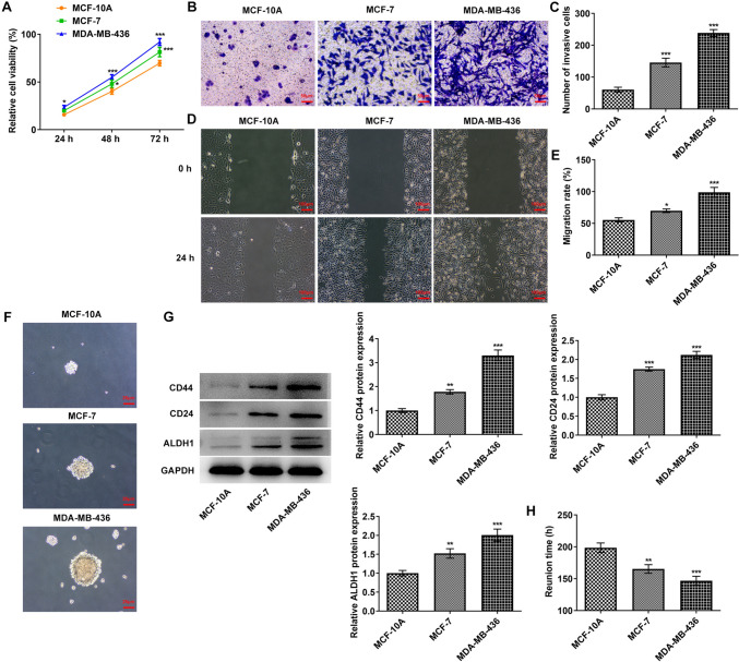
Fig. 1.
Tumor sphere formation of MCF-10A, MCF-7 and MDA-MB-436 cells. A A CCK8 kit was used to detect the cell proliferative capacity. B Transwell detected the cell invasion ability. C Statistical analysis of cell invasion ability. D Wound healing detected the cell migration ability. E. Statistical analysis of cell migration ability. F Tumor sphere formation of each group was detected by tumor sphere formation assay. G Western blot detected the expressions of CD44, CD24 and ALDH1. H Statistical analysis of the time of tumor sphere formation. *p < 0.05, **p < 0.01, ***p < 0.001 vs MCF-10A

