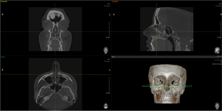
Fig. 8.
Computed tomography of the nasal cavity prior to CT-assisted precision rhinoplasty, depicting the distortions of the narrow internal nasal valves, hypertrophic turbinates, soft tissues thickening of sidewalls and septum, dimensions and relationships between chondral septum, ethmoid bone, and vomer; 3D reconstruction is extremely useful for precise understanding of anatomy for individualized surgical plan and predictable maneuvers focused on specific nuances

