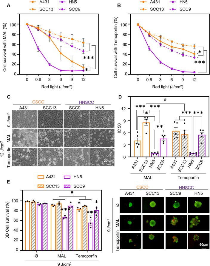
Figure 4.
Photodynamic therapy with methyl-aminolevulinate and Temoporfin. Cell survival was determined by MTT assay 24 h after incubation with 0.5 mM MAL for 5 h (A) or 24 h with 25 nM Temoporfin (B) and subsequent irradiation with red light (0 to 12 J/cm2). The results of the MTT assay are relativized to the values of absorbance at 542 nm obtained for untreated cells (indicated as control), n = 5. (C) Cell morphology after PDT (5 h of MAL or 24 h Temoporfin incubation followed by 12 J/cm2 dose) and observed by phase contrast microscopy 24 h after irradiation. (D) Half maximal inhibitory concentration (IC50) is represented for both treatments in each cell line, n = 5. (E) Cell survival after PDT (0.5 mM MAL, 9 J/cm2 or 25 nM Temoporfin, 9 J/cm2) in spheroids. Quantification of cell survival was determined by staining with acridine orange and propidium iodide and estimating the green (live) cells with respect to red (dead) cells, n = 3. Values were represented as mean ± SEM (*p < 0.05, **p < 0.01, ***p < 0.001, #p < 0.05 MAL vs Temoporfin).

