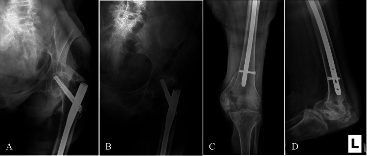
Figure 6. Six-week postoperative radiographs of the left femur.
A) Anteroposterior view of the left proximal femur demonstrating mild collapse of the fracture and 1 mm of screw cut-out. B) Lateral view of the left proximal femur. C) Anteroposterior view of the left distal femur. D) Lateral view of the left distal femur.

