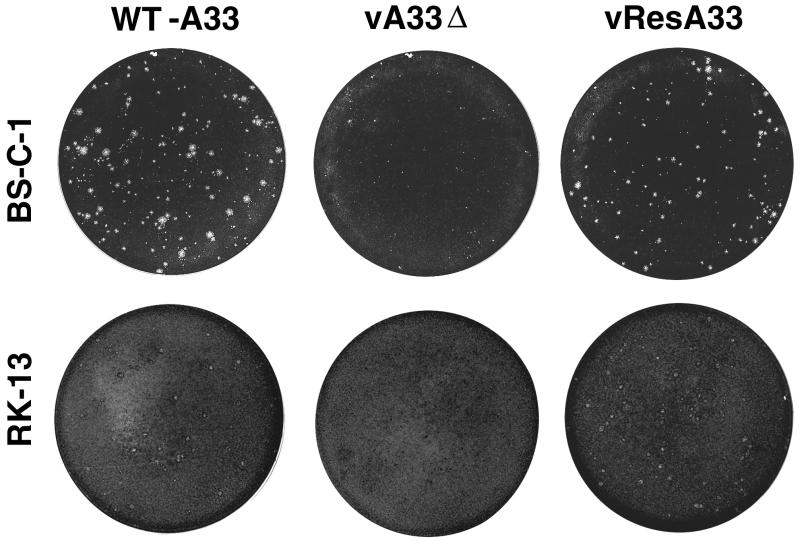
FIG. 4.
Appearance of plaques formed by the A33R deletion mutant. BS-C-1 or RK13 monolayers were infected with WT-A33, vA33Δ, or vResA33. After 48 h, the medium was removed and the monolayers were fixed and stained with 0.1% crystal violet in 20% ethanol. At least 30 infectious foci or plaques are present in each well shown.

