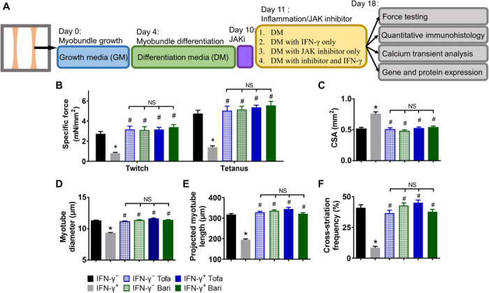
Fig. 5. JAK inhibitors prevent IFN-γ–induced deficits in myobundle force generation and structural organization.

(A) Schematic overview of myobundle culture, treatment, and characterization. Human primary myoblasts were expanded in 2D culture and mixed with hydrogel to form 3D myobundles, which were cultured in GM for 4 days, then in DM for 6 days, after which JAK inhibitors (JAKi) tofacitinib (Tofa, 500 nM) or baricitinib (Bari, 500 nM) was applied for additional 8 days, the last 7 of which in the presence or absence of IFN-γ. (B) Contractile force amplitude per myobundle CSA (specific force, n = 11 to 15 myobundles from three donors per group). (C) Quantified CSA of myobundles. (D) Quantified myotube diameter from myobundle cross sections (n ≥ 720 myotubes from three donors per group). (E and F) Quantified (E) projected myotube length (n > 150 myotubes from two donors per group) and (F) SAA cross-striation frequency (n ≥ 30 images from three donors per group) from longitudinal sections of myobundles. *P < 0.05 versus IFN-γ−, #P < 0.05 versus IFN-γ+. NS, nonsignificant. Data are presented as means ± SEM.
