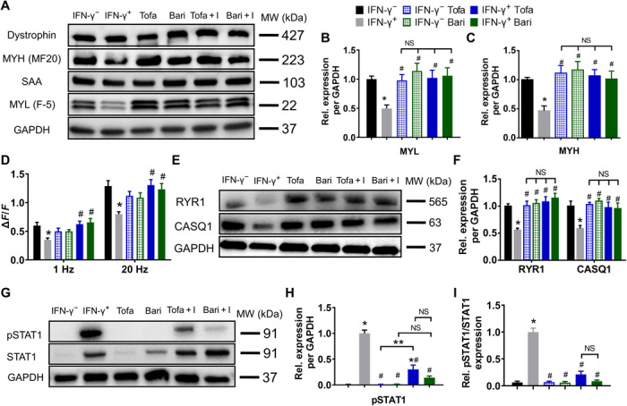
Fig. 6. JAK inhibitors prevent IFN-γ–induced protein down-regulation and STAT1 activation in myobundles.

(A) Representative Western blots from a single donor showing expression of dystrophin, MYH (MF20), SAA, and MYL (F-5), with GAPDH serving as a protein loading control. Tofa + I: tofacitinib + IFN-γ, Bari + I: baricitinib + IFN-γ. (B and C) Quantifications of Western blots for (B) MYL and (C) MYH averaged for three donors with protein abundance normalized to GAPDH and shown relative to IFN-γ− group (n = 6 samples from three donors per group). No difference in dystrophin or SAA expression was observed. (D) Quantified amplitudes of electrically stimulated (1 and 20 Hz) Ca2+ transients (n = 8 myobundles from one donor per group). (E) Representative Western blots for RYR1, CASQ1, and GAPDH. (F) Quantified RYR1 and CASQ1 protein expression normalized to that of GAPDH and shown relative to the IFN-γ− group (n = 6 samples from three donors per group). (G) Representative Western blots for pSTAT1, STAT1, and GAPDH. (H and I) Quantified (H) pSTAT1 and (I) pSTAT1/STAT1 protein expression normalized to that of GAPDH and shown relative to the IFN-γ+ group (n = 3 samples from three donors per group). *P < 0.05 versus IFN-γ−, #P < 0.05 versus IFN-γ+; **P < 0.05. NS, nonsignificant. Data are presented as means ± SEM.
