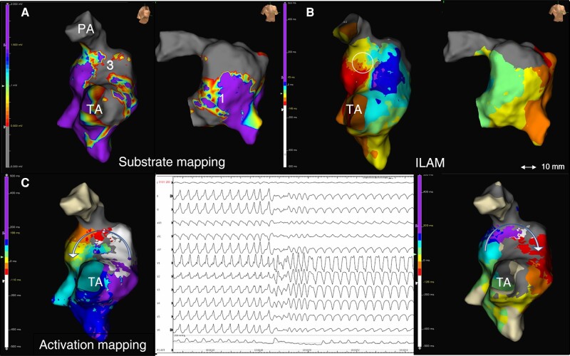
Figure 2.
Proposed working method. 3D-EAM of Patient #7: non-contemporary rTF with large transannular patch. (A) Substrate mapping showing a narrow AI3 and a large AI1 (right lateral and RAO views). (B) ILAM reveals an isolated DZ in AI3 (circle). (C) A well-tolerated VT was induced that changes during activation mapping (central panel). Activation mapping of both VT morphologies was possible in a short time using HDGC, revealing the same critical isthmus at AI3 with reverse change in wave-front propagation.

