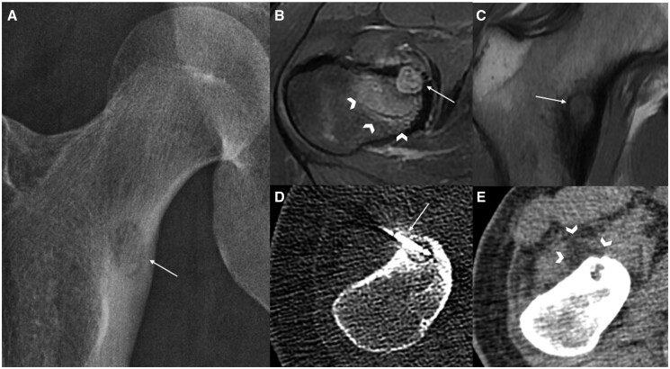
Figure 6.
Cryoablation for osteoid osteoma in a 20-year-old patient. (A) Anteroposterior hip radiograph revealed a small lucent lesion with a narrow zone of transition in the basicervical region of the right femoral neck (arrow). (B) MRI axial T2W fat-suppressed image revealed a well-circumscribed cortical-based T2W hyperintense lesion (arrow) and adjacent marrow oedema (arrowheads). (C) MRI coronal T1W image revealed a well-circumscribed lesion isointense to muscle (arrow). Findings are consistent with an osteoid osteoma and decision was made to treat via cryoablation. (D) Under CT guidance, an IceSphere ablation probe (Boston Scientific, USA) was advanced into the lesion (arrow). (E) Intra-procedural CT in soft tissue window confirmed ice formation (arrowheads) encompassing the lesion.

