Fig. 4.
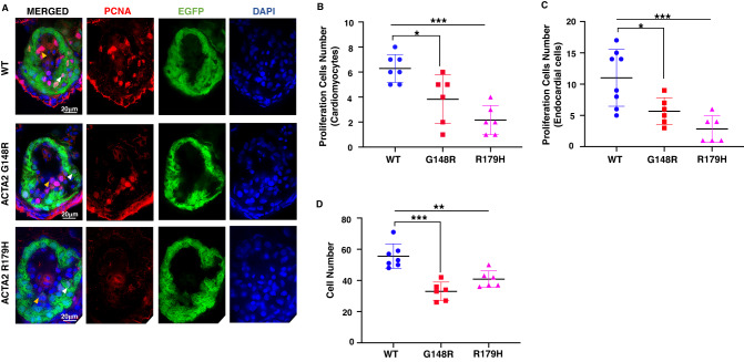
Proliferating cells were reduced in the ACTA2 G148R variant. A–D Immunostaining of Tg[cmlc2:EGFP] ACTA2 larvae heart at 4 days post-fertilization (green) with proliferation cell marker (PCNA, red), 4’,6-diamidino-2-phenylindole (DAPI) co-staining (blue). Scale bar: 20 μm. A Endocardial cells (yellow arrowheads) and cardiomyocytes (white arrowheads). Zebrafish with ACTA2 p.G148R and ACTA2 p.R179H show reduced total cell numbers. B In the myocardium, ACTA2 p.G148R and ACTA2 p.R179H cardiomyocyte showed decreased numbers of proliferating cells in the heart wall area highlighted with EGFP green fluorescence, PCNA, and DAPI. C In the endocardium area, endocardium cells showed decreased proliferation in all pathogenic variants highlighted with PCNA and DAPI. **P < 0.01, ***P < 0.001. Error bars indicate the standard deviation

