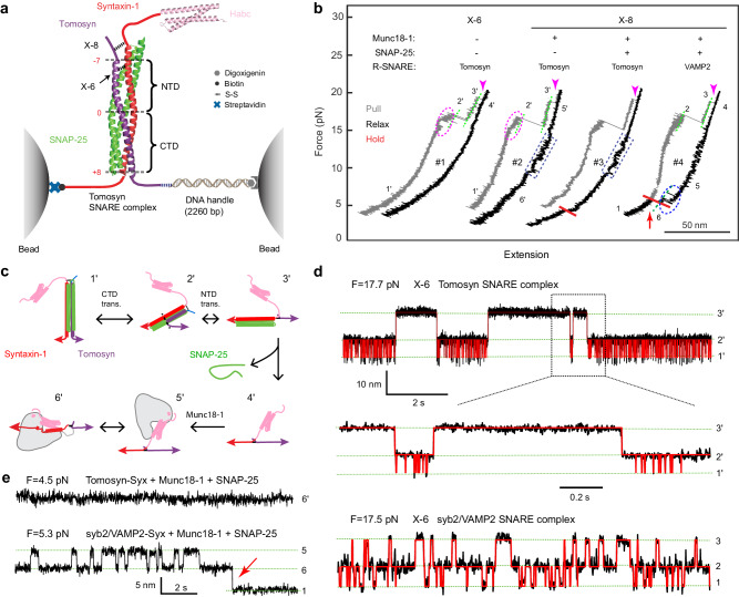
Fig. 4. The SNARE motif of tomosyns forms a stable SNARE complex with syntaxin-1 and SNAP-25, but fails to form a template complex with Munc18-1 and syntaxin-1.
a Experimental setup to pull a single tomosyn SNARE complex (PDB ID: 1URQ) using optical tweezers. Tomosyn and syntaxin-1 (Syx) molecules in the pre-assembled complex were attached to two polystyrene beads via a DNA handle at their C-termini and crosslinked at either the -6 layer (X-6) or the −8 layer (X-8). The central ion layer divides the parallel helix bundle into the N-terminal domain (NTD) and the C-terminal domain (CTD). Munc18-1 and SNAP-25 may be added to the solution either alone or together to test template complex formation or SNARE assembly. The presence or absence of these proteins free in solution is indicated by “+” or “−“, respectively. b Force-extension curves (FECs) obtained by pulling (gray) or relaxing (black) the tomosyn-Syx conjugate at a speed of 10 nm/s or holding it at a constant mean force or trap separation (red). Different FEC regions, indicated by green dashed lines, are labeled by the numbers of their associated states in panel c. The dashed magenta oval, blue rectangles and blue oval mark reversible folding and unfolding transitions of tomosyn SNARE four-helix bundle (see Fig. 4d), Munc18-1-bound open syntaxin and the template complex, respectively. Red arrows indicate events of SNAP-25 binding to the template complex. Magenta arrow heads mark the unfolding of t-SNARE complexes and the accompanying dissociation of SNAP-25 from the pre-assembled SNARE complexes. c Schematic diagrams of different states involved in SNARE folding/assembly and Munc18-1 association, including the fully assembled tomosyn SNARE complex (1’), the half-zippered four-helix bundle (2’), the unzipped tomosyn (3’), the unfolded SNARE motifs (4’), the unfolded SNARE motifs with Munc18-1 bound to the N-terminal region of syntaxin-1 (5’), and the Munc18-1-bound open syntaxin (6’). See Fig. 5c for similar states with tomosyn replaced by VAMP2. d Extension-time trajectories at the indicated constant forces (F) showing three-state folding and unfolding transitions of the tomosyn or syb2/VAMP2 SNARE complex. Red curves represent the idealized transitions derived from hidden Markov modeling. Green dashed lines mark the average extensions of the associated states labeled on the right. e Extension-time trajectories of different SNARE conjugates at constant low forces.

