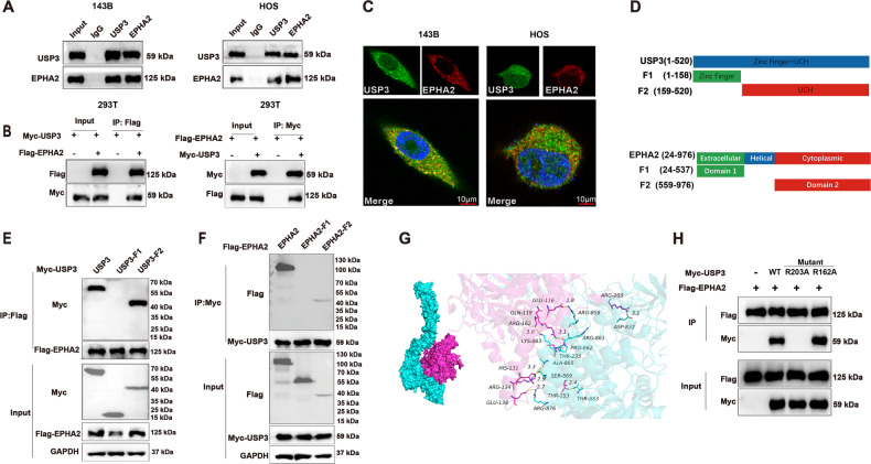
Fig. 4. USP3 interacts with EPHA2.
A Cell lysates were immunoprecipitated with control IgG, anti-USP3, or anti-EPHA2 antibodies. The precipitates were detected by WB. B HEK293T cells transfected with the indicated plasmids were immunoprecipitated with anti-Flag or anti-Myc antibodies. The lysates and precipitates were analyzed. C 143B and HOS cells were double-stained for USP3 and EPHA2 and observed by confocal microscopy, and the nuclei were counterstained with DAPI. (Scale bars, 10 µm) D Overview of USP3 and EPHA2 structures. E, F HEK293T cells transfected with the indicated constructs were immunoprecipitated with anti-Flag or anti-Myc antibodies. The lysates and precipitates were detected. G Molecular model of docking between USP3 (purple) and EPHA2 (green). H Mutations of R162A and R203A were introduced into wild-type (WT) USP3 with a Myc tag, and Co-IP was then performed to examine the interaction of these mutants with EPHA2. R162A: the arginine at position 162 was replaced by alanine; R203A: the arginine at position 203 was replaced by alanine.

